A Bone Density Scan is a safe, quick and painless exam that helps diagnose osteoporosis and assess the risk of fractures, among other things. This scan is especially recommended for people over the age of 40, due to the natural loss of bone density that occurs with age. Find out how this exam is performed, when it should be done and what precautions to take.
What is a Bone Density Scan?
A bone density scan is an imaging examination that measures the bone density and mineral content in different parts of the body, using low-dose X-ray. In other words, this scan is used to quantify bone loss, which occurs naturally with age, and is therefore the best exam to diagnose osteoporosis.
This scan is performed on the spinal column, wrist or hip, and the results obtained can be compared with one of two scores. The T-Score is a comparison with a healthy young adult at the peak of their bone density, while the Z-Score compares the results of this scan with a healthy person of the same age and body size.
What is a Bone Density Scan for?
A bone density scan serves to determine the early stages of bone loss associated with osteoporosis; thus, it is essential in the fight against this disease. Based on the results, it is possible to assess the risk of developing fractures and devise an effective therapeutic strategy to prevent osteoporosis.
Furthermore, bone density scans also help verify the effectiveness of osteoporosis treatments. Therefore, this is a way of monitoring therapy, allowing treatment to be adjusted according to the progress observed, also taking into consideration other important factors, such as age, body weight and risk behaviours, like smoking or excessive alcohol consumption.
Bone Density Scan: how is it done?
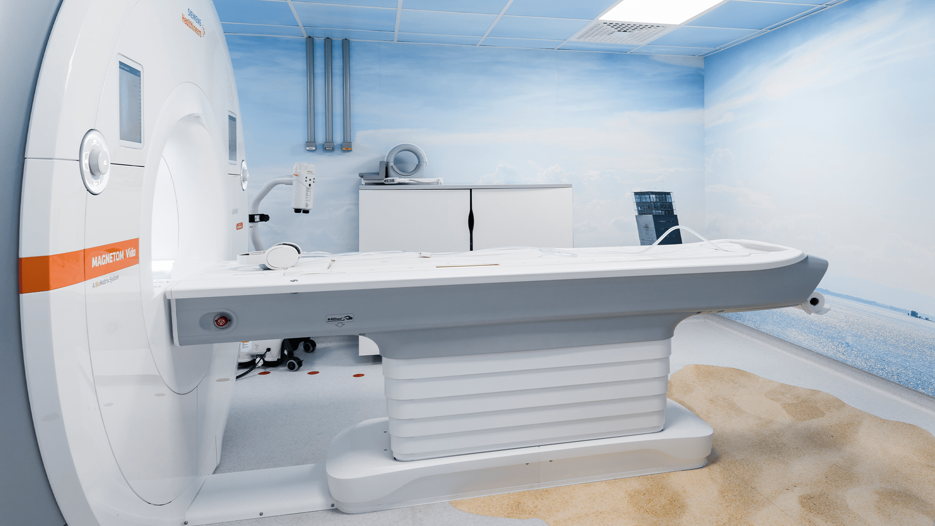
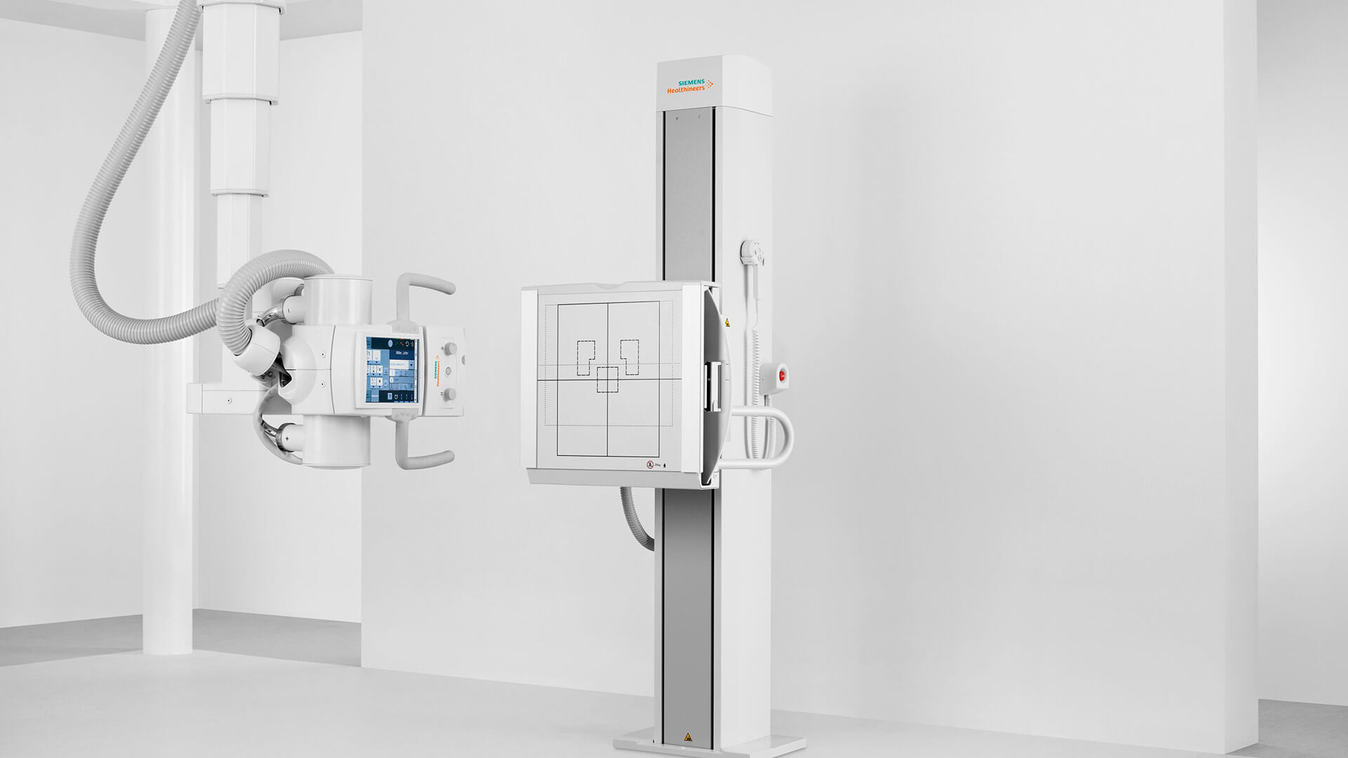
A bone density scan is performed using equipment that measures bone density in the whole body, such as the spinal column, hip and wrist.
To begin the exam, the patient lies on a table above an X-ray generator, while a detector slowly passes over the area in question to collect the image. During the scan, the patient should remain still to avoid distorting the image generated. It may be necessary to hold their breath for a few seconds to obtain a more precise image. On average, the exam takes 15 to 20 minutes, depending on the areas analysed.
After the scan, a specific algorithm calculates the patient’s bone density and compares the results with reference scores, generating a graph. The medical specialist interprets the results and assesses the need for therapeutic intervention.
Who should undergo a Bone Density Scan?
According to recommendations by the World Health Organization (WHO), bone density should be measured as a routine test for women over the age of 65 and men over .
Furthermore, this exam should be performed on anyone, of any age (including children), if they present one of the following risk factors:
• Personal or family history of vertebral fractures;
• Low body mass index;
• Takes corticosteroids, anticonvulsants or certain barbiturates;
• Frequent falls;
• Excessive alcohol consumption;
• Smoking;
• Thyroid disorders;
• Type-1 diabetes, liver and kidney disorders.
In addition, bone density scans should be repeated every 2 years to monitor changes in bone density, whether increases or decreases. You should always consult your physician to determine the need and frequency for this screening in your specific case, and follow all instructions.
What to expect from a Bone Density Scan?
The bone density results are interpreted by the medical specialist and can be compared against two reference scores: the T-Score or the Z-Score. The first is the most widely used, as it helps estimate the patient’s risk of suffering a fracture and determine the most appropriate course of treatment.
Therefore, the scores can be placed in the following groups:
• T-Score equal to or above -1,0: normal bone density, without risk of fracture;
• T-Score between -1,0 and -2,5: early stage loss of bone mineral density (called osteopenia), when there is already a risk of fracture;
• T-Score equal to or below -2,5: significant loss of bone mineral density and high risk of fracture.
What are the restrictions of a Bone Density Scan?
While bone density scans are essential to measure bone density, they can only indicate the relative risk and cannot predict when a patient will suffer a fracture. Furthermore, it may be of little use in patients with spinal deformities or who have had surgery in this area, as these factors interfere with the quality of the image obtained. In these cases, a computerised tomography (CT scan) is more effective.
It is also important to note that follow-up exams must be performed in the same area and by the same equipment. Different machines have different sensitivities and may take measurements with some degree of deviation. Therefore, it is not possible to directly compare two bone density scans obtained from different machines.
What precautions must be taken with a Bone Density Scan?
Bone density scans are quick, painless and safe. Despite the use of ionising radiation, the dose emitted during the exam is very low, indeed, much lower than an X-ray, representing less than one day of sun exposure.
However, pregnant women should always inform their physician before undergoing the exam. If the physician recommends a bone density scan, it may be necessary to take measures to protect the foetus from exposure to radiation.
In addition, patients should inform the physician if they have undergone any prior exams, especially those using contrast media. In some cases, the patient may have to wait a few days before undergoing this scan.
Patients can maintain their regular diet, but should not take calcium or magnesium supplements for 24 hours before the exam. It is advisable to wear comfortable clothing and avoid metal accessories or details.
Bone Density Scan at Joaquim Chaves Saúde
The Joaquim Chaves Saúde Imaging units are equipped with the latest technology to perform an extensive and comprehensive range of imaging examinations, with the highest quality and precision. If you need a bone density scan, don’t delay any longer. Schedule your scan now and get the results you need, precisely, quickly and safely.
