Soft tissue ultrasound is a diagnostic tool specifically designed to evaluate superficial tissues. It has broad applications across various medical specialties and offers a simple, comfortable, and immediate way to visualize structures. Learn how this examination is performed and the cases where it is indicated.
What is soft tissue ultrasound?
Soft tissue ultrasound (or ultrasound of soft tissues) is an imaging examination that allows the evaluation of superficial tissues such as the skin, subcutaneous fat, joint structures, muscles, tendons, and ligaments. This examination can diagnose traumatic and inflammatory pathologies, enabling the assessment of damages caused by trauma or abscesses. It can also be used to visualize skin changes, such as cutaneous nodules, cysts, lipomas, and other superficial lesions. Soft tissue ultrasound is commonly used to diagnose abdominal wall hernias (inguinal and umbilical hernias).
Soft tissue ultrasound does not involve radiation, making it a safe and non-invasive examination. It does not cause any pain or discomfort, and it is well-tolerated by patients of all ages and clinical conditions.
What is soft tissue ultrasound used for?
Soft tissue ultrasound has diverse and wide-ranging applications. It can evaluate diseases and disorders of superficial organs like the skin, muscles, and tendons. Additionally, it provides an initial approach to other organs such as the breasts, testicles, and thyroid, although there are specific ultrasounds for these cases. Therefore, soft tissue ultrasound plays a vital role in reaching a diagnosis and guiding the most appropriate therapeutic approach for each case.
How is soft tissue ultrasound performed?
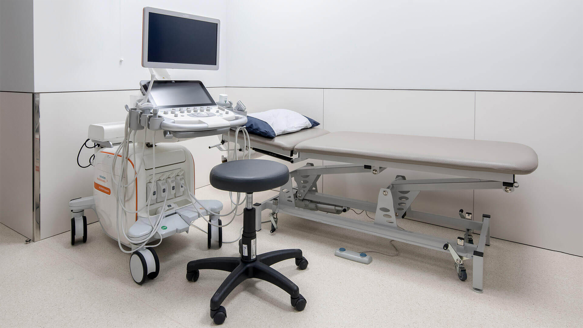
Soft tissue ultrasound uses a device called an ultrasound scanner, which captures images of the structures under study by emitting and receiving ultrasound waves. A conducting gel is applied to the area being examined, and the physician gently moves a probe over the skin to direct the sound waves and obtain internal images.
The obtained images are sent to a monitor, where they can be immediately visualized. The physician selects, analyzes, and interprets the most important and representative images. If no abnormalities are observed in the structures, the soft tissue ultrasound is considered normal. On the other hand, if the images show changes, they may be sufficient for a diagnosis or indicate the need for further tests to clarify the ultrasound findings. Soft tissue ultrasound is a quick examination with an average duration of 5 minutes. It may take longer if a more detailed observation of the anatomical area under study is required.
What precautions should be taken for soft tissue ultrasound?
Soft tissue ultrasound does not require any specific preparation. Patients can eat, drink, and take their regular medication as usual. However, it is recommended to wear comfortable clothing that facilitates access to the body area being examined. Any accessories that may interfere with image quality, such as earrings, necklaces, rings, bracelets, watches, and piercings, should be removed.
What are the limitations and risks of soft tissue ultrasound?
Since soft tissue ultrasound does not involve any form of radiation, it can be safely and comfortably performed on both adults and children, including infants. It is a commonly requested examination in pediatrics due to its painless, comfortable, safe, and effective nature. Moreover, pregnant women can undergo soft tissue ultrasound, unlike other examinations that involve radiation (such as computed tomography - CT scan and conventional X-ray).
Soft tissue ultrasound at Joaquim Chaves Saúde
The Imaging Service at Joaquim Chaves Saúde is equipped with state-of-the-art diagnostic imaging tools operated by an experienced and specialized team. Benefit from cutting-edge technology to obtain the results you need with maximum speed, precision, safety, and comfort. If you need to undergo a soft tissue ultrasound, schedule your examination with us.
Explore here other types of available ultrasounds.


